-
SPECIALISATION ABROAD
Till end of March 2023, I will work three weeks per month in Finland.
One week monthly I'll be in Bulgaria and able to perform planned surgeries. For this purpose, initial diagnostics, performed by the referring veterinary surgeon is necessary first.
As an exception, I may do some examinations and consultations, especially in complicated cases and as a second opinion.
For more information, please, write to: (Please enable JavaScript to view this email address)
Dr. Vladislav Zlatinov
Graduated as Master of Veterinary Medicine in 2005 in Sofia.
From 2005 to 2009 worked as a general practitioner at the Animo Small Animal Veterinary Clinic.
Since 2009 working as a chief veterinarian and senior surgeon at the Central Veterinary Clinic - Sofia, gaining main clinical experience and became a specialist in the field of orthopedics and spinal surgery.
In the last 5 years, the main focus of my work has been referred surgical cases, requiring complex diagnostic and therapeutic activities.
From 2021 started working as a freelance surgeon.
Memberships and recognitions
2012: co-founder of the Bulgarian Association of Veterinary Orthopedics and Traumatology (BAVOT). Currently President of the Organization.
2013: certified member of GRSK - FCI recognised organization for control of genetic diseases of the skeleton (internationally certified panelist for elbow and hip dysplasia).
2015: co-founder of Veterinary Orthopedic and Neurology Group (VOG) - Slovenia.
2015: AOVET - active member, speaker / instructor (AOvet faculty).
Practical trainings and certificates
2007: two-week externship in the surgical department of the Clinic Mediterraneo, Madrid, Spain, under the direction of Dr. Miguel Ruiz Fundador and Dr. Margot Ruiz - general orthopedic and neurosurgery cases.
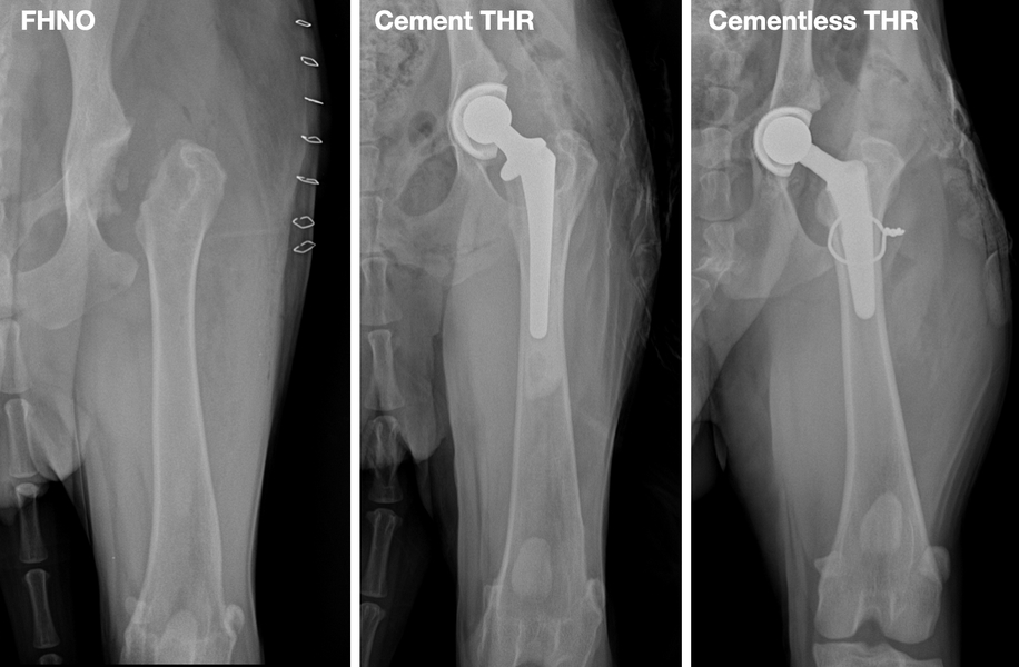
2012: course and certification program for Total hip replacement - BIOMEDTRIX Universal Hip - Bristol, UK.
2014: two-week internship at Clinica Parabiago, Milan, Italy, under the guidance of Dr. Luca Redelli. Training with emphasis on TPLO technique for ruptured cranial cruciate ligament and distraction osteogenesis (Ilizarov’s method).
2014 AOVET courses:
- Principles in a Fracture management; Zagreb, Croatia;
- Advances in Fracture management; Cremona, Italy;
- Master class on advanced corrective osteotomies: Rear limb and Patellar luxation, Cremona, Italy.
2014: one-week externship at the Faculty of Veterinary Medicine, Turin, Italy, under the guidance of Professor Bruno Peirone. Training with focus - traumatology, TPLO technique for ruptured cranial cruciate ligament.
2015: conducts 8 weeks AOvet fellowship internship at Clinica Veterinaria - Dr. Aldo Vezzoni, Cremona, Italy, with emphasis on diagnosis and treatment of hip and elbow dysplasia, total hip arthroplasty (Kyon THR), TPLO, arthroscopic diagnosis and treatment.
AOvet fellowship: a sponsored postgraduate program in orthopedics.
Speaker / instructor in regional seminars
Frequent speaker in national seminars - BAVOT and others.
Instructor of practical courses (wet lab) - X-ray examination of the skeleton, Surgery of the stifle joint, External skeletal fixator for the treatment of fractures held in Central Veterinary Clinic - Sofia.
“Osteosynthesis with plates” (dry lab), Mikromed sponsored seminar, Sofia.
TPLO technique for ruptured cranial cruciate ligament (International wet lab), VetWest, Sofia.
International speaker / instructor
“Advanced Orthopedic Workshop” - Opatija, Croatia, 2016.
“Juvenile orthopedic diseases”, VOG - Zagreb, Croatia, 2017.
“Sankt Petersburg International Vet Conference” (Russia), 2016 and 2017.
“CORA based TPLO”, practical course, St. Petersburg, 2017.
Fracture treatment principles, Limassol, Cyprus 2017.
Main speaker in the orthopedic section of the National Veterinary Conference - Moscow, Russia, 2018.
“Traumatology Seminar” - VOG, Zreče, Slovenia, 2018.
“Fracture treatment by plate osteosynthesis”, practical course, Bucharest, 2018.
Lecturer in the orthopedic section of CONGRESO INTERNACIONALVETERINARIO DE LEON- Leon, Mexico, 2019.
International lecturer / instructor Intrauma (Fixin) courses
“FIXIN wet lab on TPLO and DPO” - co-lecturer with Dr. Luca Vezzoni - practica lcourse, Opatija, Croatia, 2017.
“TPLO dry lab-basic course” - co-lecturer with Dr. Massimo Petazzoni, practical course, Bucharest, Romania, 2017.
Fixin international symposium - case presenter at round table discussion, Venice, 2017.
“Angular bone deformities - hind limb” - co-lecturer together with Dr. Luca Vezzoni, practical course, Opatija, Croatia, 2018.
“TPLO dry lab” - co-lecturer together with Dr. Luca Omodeo, practical course, Larnaca, Cyprus, 2019.
“Second Intrauma Symposium” - speaker, Turin, Italy 2019
AOVET faculty / table instructor
"Small Animal Master Class - Small and Toy Breed Dog" - Dubrovnik, 2016.
“Masters in Small Animal - Tibial Plateau Levelling Osteotomy (TPLO)” - Lisbon, Portugal 2017.
“Canine Sports Medicine Orthopedics and Traumatology”, master course, Dubrovnik, 2018.
“Avian and Exotics Orthopedics”, master course, Dubrovnik, 2018.
Asian Veterinary Orthopedic and Neurological Conference - Xian, China, 2019
Three days courses and lectures:
- Dynamic Ulna Osteotomies and Proximal Abducting Ulnar Osteotomy (PAUL), Patellar Groove Replacement (PGR) for Patellar Luxation
- AO VET Surgical Secrets
- Complex Cases and Advanced Surgery
Other
Co-organizer in the planning and conducting of over 10 seminars with international lecturers of the Bulgarian Association of Veterinary Orthopedics and Traumatology (BAVOT).
Author of 15 publications in regional scientific journals (including online publications) - articles and clinical reports.
One of the first veterinary surgeons to introduce in Bulgaria - total hip replacement, TPLO technique for ruptured cranial cruciate ligament, arthroscopic method for treatment of orthopedic diseases and others.
The first surgeon in Bulgaria to introduce transcutaneous integrated limb endoprosthesis (still a highly innovative technique worldwide).
AO VET Dubrovnik, 2017
AO VET Lisbon, 2019
BAVOT, 2019, corrective osteotomy of the hindlimb
BAVOT, seminar with Mike Farrell and Ignacio Calvo, 2018
BAVOT, seminar with Prof. Bruno Peyrone
Intrauma symposium, 2019
Leon Congress, Mexico, 2019
AO VET, Pan-Asian Conference, 2019
TPLO workshop with Massimo Petazzoni, 2017
VOG seminar, Croatia, 2016
I have many years of experience in the medicine of small
animals.
For over a decade I work mainly in the field of veterinary orthopedics and neurosurgery.
In these areas I am fully competent in the whole treatment process - from the clinical examination to the postoperative rehabilitation.
My orthopedic and neurosurgical techniques are in accordance with the latest world standards.
I have extensive experience, as well, in soft tissue surgery - abdominal, thoracic and reconstructive.
For patients
As a freelance surgeon, I am a specialist in the diagnosis and treatment of surgical diseases.
The way I work requires that the consulted patients are not emergency (i.e. to permit planned work).
I perform many types of orthopedic, neurosurgical and soft tissue operations, as well as, consultations and treatment in other fields like oncology, upper respiratory tract diseases (e.g. brachycephalic syndrome) and plastic reconstruction of soft tissue defects.
For additional details about consultation and appointments, please see here.
For veterinarians
I offer collaboration in the diagnosis and treatment of surgical diseases, expertise and systematic approach to the patients.
My main field of expertise is orthopedics and neurosurgery, but I also have extensive experience in abdominal and thoracic surgery.
I do clinical and specialised examinations of patients with orthopedic and spinal diseases, as well as vast numbers of surgical interventions.
Here, you can see details and a list of the performed diagnostic and surgical procedures.
Title | Author |
---|---|
Patellar luxation | Dr Vladislav Zlatinov |
Intervertebral disc disease | Dr V. Zlatinov |
Cranial cruciate ligament disease / rupture | Dr V. Zlatinov |
Additional details for veterinarians | Dr V. Zlatinov |
Additional details for patients | Dr V. Zlatinov |
More about me | Dr V. Zlatinov |
What is patellar luxation?
The patella (knee cap) is a small bone located at the base of the quadriceps muscle tendon. Its function is to act as a fulcrum in the movement of the knee joint. When bent and stretched, it slides into a special sulcus (trochlea) on the front part of the femur bone.
In some dogs, the patella is displaced (luxated) from this normal route, which adversely affects the proper function of the joint. As a consequence the altered biomechanics, some patients develop varying degrees of lameness and osteoarthritis in the long term.
How can I tell if my pet has patellar luxation?
Patellar luxation is a common condition. It most often affects dogs (rarely cats) of small breeds. Clinical signs appear most often at an early age of 4-10 months. A characteristic "skipping" lameness is often observed, in which the animals limp for a few steps and then quickly return to normal. Other animals are lame all the time (large breeds), and when both knees are affected, the dogs have a stiff posture with the weight transferred to the front of the body.
What is the cause of patellar luxation?
The reasons for the development of pathological conformation of the limbs are genetic. In fact, most animals are born with normal joints, but early in life they develop abnormalities in the of muscles and tendons.
Most often the deviation of the patella is to the medial (internal) aspect of the knee joint. When the mechanism of the quadriceps muscle is displaced in this direction, it acts as a "bowstring" and causes deformity of the growth of the bones of the limb. The trochlea, in which the patella is to be placed, does not develop properly and is sometimes even completely missing.
What could be the consequences for the affected joint?
In case of luxation of the patella on the joint surface, damage to the articular cartilage is possible. This is one of the reasons for more permanent pain and potentially progressive osteoarthritis. Also, the abnormal direction of traction of the thigh muscle causes internal rotation of the tibia relative to the femur, which can strain the cranial cruciate ligament. This is why it is common for patients with medial patellar luxation to eventually damage their cruciate ligament further in life.
How is patellar luxation diagnosed?
Patellar luxation is usually diagnosed during a clinical examination by an orthopaedic specialist.
To assess the bone deformities leading to patellar luxation, it is necessary to perform specific radiographic projections. To avoid false artifacts, it is extremely important to position the patient correctly, which is only possible with the use of sedation (superficial anesthesia).
Less often, computed tomography (scanner) is required for assessment of the condition in complex cases.
What are the degrees of patellar luxation?
The classification of patellar luxation is based on the mobility of the cap relative to the femoral trochlea:
Grade 1: The patella can be luxated under pressure, but returns immediately to its place;
Grade 2: The patella is displaced spontaneously in flexion and extension of the knee, but also spontaneously reduces itself;
Grade 3: The patella is permanently luxated, but can be reduced under pressure;
Grade 4: The patella is permanently luxated and cannot be reduced.
How is patellar luxation treated?
Sometimes patellar luxation is found accidentally during a routine clinical examination. In adult dogs, this accidental finding suggests a conservative approach.
In young animals of small breeds, treatment depends on the degree of luxation and clinical signs.
The conservative approach is appropriate for grade 1 and 2, without clinical signs (lameness). In higher degrees (3 and 4) and in all clinically manifested cases, surgical treatment is required.
Large breeds of dogs always require surgical treatment, regardless of the degree of luxation.
What is the non-surgical treatment of patellar luxation?
In fact, there is no real non-surgical treatment for the problem. Some measures, such as weight control, physiotherapy and painkillers, can help control clinical signs.
Surgical treatment of patellar luxation
Surgery is recommended in dogs with lameness as a result of patellar luxation.
There are many surgical techniques. Their main purpose is to restore the normal direction of traction of the quadriceps muscle relative to the skeleton of the limb. This requires correction of bone, cartilage and soft tissue structures.
There are two groups of techniques: Classic (simple) techniques and more invasive corrective osteotomies.
In fact, a single surgical technique is rarely adequate enough, often a combination of methods is required.
Classic techniques (simple) - are applied independently in mild degrees of the disease, when there are no serious bone deformities.
Transposition of the tibial ridge.
After a partial osteotomy, a small area of the tibia that terminates the patellar tendon is moved inward or outward to correct the direction of muscle tension.
The fixation is with small pins; healing takes about 4 weeks.
Trochleoplasty
When the femoral trochlea is shallow, it may be necessary to deepen the sulcus. This includes a special remodeling of the bone and cartilage wedge, which deepens its position.
Reconstruction of soft tissues
Often the soft tissues on both sides of the patella are either too tight or too loose. Reconstructions are usually performed to release tight tissues and tighten (suture) loose tissues.
Corrective osteotomies
In some dogs (mostly large breeds) with high-grade patellar luxation, the deformity of the femur or the tibia are very important causative factors.
To decreases the risk of recurrence, in certain cases it is strongly indicated to correct the bone by a procedure, called osteotomy.
This is a more invasive surgery in which the bone is cut and a bone wedge is removed, and the new shape is fixed with a plate and screws. B
one healing in these cases usually takes about 6 weeks.
Patellar Grove replacement
In rare cases of severe arthritis affecting the femoral trochlea, a partial joint replacement is indicated. For this purpose, a commercial PGR prosthesis with a very smooth (diamond) surface is used, which replaces the altered cartilage structure.
Will my dog recover fully after the operation?
Your dog's physical activity should be strictly limited for a period of about 6 weeks. This is a period in which the tissues in the area heal and the knee joint adapts to the new biomechanics.
Overloading the limb at this time can lead to complications and slower recovery.
In certain cases, during this period, physiotherapy may be indicated to speed the recovery.
After the 6-week postoperative period, a follow-up examination is needed to confirm the healing of the tissues around the knee. Upon confirmation, the dog can return to normal physical activity without restrictions.
Is the congenital patellar luxation different?
This is the most severe form of the disease, in which the patient is born with luxated patellae. Some authors call this form grade 5 because the severity of bone deformities is unusually advanced.
With delayed diagnosis, distorted bone structures, shortened muscles and ligaments make treatment extremely challenging. Unfortunately sometimes normal clinical recovery is impossible.
If you notice a strange gait and conformation of the limbs in young puppies, it is necessary to consult a veterinarian as soon as possible in order to avoid delays in treatment.
CONCLUSION
https://doctorzlatinov.com/EN/#article-31
What is intervertebral disc disease?
Intervertebral disc disease is the most common disease of the spine in dogs (much less common in cats).
Intervertebral discs are fibrocartilage structures between the vertebrae that allow micromovement and act as shock absorbers. They consist of a fibrous outer ring (anulus), and jelly-like core (nucleus).
OpenStax College, CC BY 3.0, via Wikimedia Commons
Degeneration of the intervertebral discs leads to changes in the histological structure that reduce their elastic properties.
The end result can be a “rupture” of disc material (disc herniation) and compression of the spinal cord.
Laboratoires Servier, CC BY-SA 3.0, via Wikimedia Commons
What causes intervertebral disc disease and which breeds are predisposed?
Some breeds (chondrodystrophoid) are genetically predisposed to intervertebral disc degeneration. In them, changes (dehydration) in the structure of the connective tissue begins at an early age
Such are young (up to 5-6 years old) small breeds of dogs as Dachshund, Basset hound, Japanese hin, lhasa apso, Shih tzu, Pekingese, Cocker spaniel, and others. The reason for this is that they were genetically selected for the exterior of “dwarf dogs” and most of these dogs have a long body and short limbs.
Unfortunately, exterior selection has led to a defect in the development of cartilage of the entire skeleton, including the intervertebral discs.
An abnormal elasticity of the discs in them, can lead to disc herniation and compression of the spinal cord.
There are three types of disc herniation- Hansen I, Hansen II и Hansen III.
Acute disc herniation (Hansen type I, extrusion)
Small breeds of dogs at a young age (between 2 and 6 years) are most often affected. Larger breeds are very rarely affected.
Clinical signs usually appear suddenly.
Disc herniation is most easily described as „extrusion“ or leakage of the internal contents of the intervertebral disc.
When the disc changes, it loses its stability and even normal movements (especially torsion) lead to its rupture. Unfortunately, this is always to the vertebral canal, therefore compressing the spinal cord.
The speed of extrusion and the volume of the ruptured disc affect the severity of the spine damage.
The severity of the clinical signs varies from pain syndrome to complete paralysis.
Severe cases of herniated disc (Hansen type I) are an emergency that requires prompt consultation with a veterinary surgeon.
"Disc Disease" (Hansen type II, protrusion)
This disease is similar to disc disease in humans. Instead of extrusion of the center of the disc, there is a prolapse (protrusion) of the periphery.
Unlike acute rupture, the symptoms usually develop more slowly and progressively. Larger breeds of dogs (German Shepherd, hunting breeds), at a later age of 5-12 years, are relatively more often affected.
Laboratoires Servier, CC BY-SA 3.0, via Wikimedia Commons
Traumatic disc herniation, Hansen type III - acute noncompressive hernia
The cause of this type of hernia is a specifically directed spinal cord trauma, which leads to an explosive loss of the normal disc nucleus in the event of a sudden traumatic rupture of the ring (annulus). A small amount of normal disc material injures the spinal cord without subsequent compression.
Clinical signs are usually paralysis and pain.
The prognosis varies, and in mild to moderate cases, improvement is possible with rehabilitation and physiotherapy without surgery.
In severe cases, myelomalacia (a necrotic process in the spinal cord) can develop, which can be fatal.
How a ruptured disc affects the spinal cord?
Figuratively, the spinal cord is very similar to a cable, which consists of thousands of small wires. When pressed, the transmission of information through the many microscopic threads is disrupted.
In a herniated disc, the nucleus is pushed up and compresses the spinal cord or the nerves that come out of it.
Pressure on the nerve root results in varying degrees of acute pain (nerve root signature), and if enough disc material has been expulsed into the canal, the spine is compressed and neurological deficit develops.
Is a sudden onset possible?
Disc degeneration occurs relatively slowly - for weeks and months, but the disc herniation itself usually develops very quickly.
Some dogs develop severe paralysis within an hour. The faster the onset, the greater is the need of emergency veterinary care.
How can I tell if my dog has a herniated disc?
The most common symptom is back or neck pain.
In addition to vocalization, common signs are unusual posture (head bowed), trembling, rapid breathing, reluctance to move, difficulty ascending and descending furniture / stairs.
In more severe cases, there may be difficulty walking, ranging from weakness to complete paralysis.
The most severe cases are those with paralysis, lack of bladder control and loss of sensation.
If the paralysis affects all four limbs, the disc hernia is in the neck area.
How to diagnose a herniated disc?
The first guideline is a history of pain, incoordination, or paralysis without a history of trauma.
Clinical examination usually reveals whether there is a neurological deficit of the spinal cord.
Another important detail is the breed of the patient. If the dog is one of the predisposed breeds, the diagnosis is even more likely.
In some cases, a simple (plain) x-ray may help localize the problem, but the results can be misleading. The normal and often the degenerated disc are X-ray-negative.
When the patient needs surgery and the plain radiography fails to detect the problem, it is necessary to use more advanced diagnostic imaging tools - contrast examination (myelography), computed tomography (scanner) or magnetic resonance imaging.
What are the degrees of neurological deficit?
First degree is characterized by mild pain and is usually self-limiting within few days.
Second degree causes moderate to severe pain that persists longer.
Third degree partial paralysis (paresis) and incoordination of movements.
A 2-year-old French bulldog with third-degree neurological deficit due to an acute disc herniation.
In grade four, there is paralysis (inability to stand up), but with deep sensitivity.
Fourth degree neurological deficit
The most severe, fifth degree is characterized by complete paralysis and lack of deep sensitivity.
Switching from one stage to another can take place within an hour or a day.
7-year-old Dachshund with fifth-degree neurological deficit.
With a strong stimulus, the dog does not turn around and does not feel anything.
Lacks deep sensitivity.
When is conservative therapy applied?
Drug treatment can be used in patients with pain syndrome or in patients with mild deficiency. Less commonly, this therapy may be successful in more severe cases.
Dogs that have lost their sense of deep pain need urgent (within hours) surgery.
The disadvantages of conservative treatment are more frequent recurrence of clinical signs and a greater chance of permanent neurological deficit.
The most important element of conservative therapy is restriction of movement (cage rest).
Restricting the patient's movements can prevent further extrusion of disc material.
The body's natural regenerative potential can lead to self-healing of the spinal cord.
When is surgery required?
Patients with neurological deficits (paresis) may need surgery (decompression) because the prognosis is statistically much better than with conservative treatment.
In general, surgical treatment leads to faster recovery and less likelihood of recurrence.
Cases with loss of deep sensitivity (paralysis) definitely require urgent surgery and have worse prognosis.
What is the success rate in treating disc herniations with or without surgery?
Degree and duration | Recovery | |
---|---|---|
Without surgery | With surgery | |
II degree up to 1 week: | 80-90 % | 90-95 % |
II degree over 1 week: | 60-70 % | 90-95 % |
III degree: | 30-40 % | 85-95 % |
IV degree up to 3 days: | < 25 % | 85-95 % |
IV degree over 3 days: | < 20 % | 60-70 % |
V degree up to 24 hours: | < 5 % | 50 % |
V degree over 24 hours: | < 5 % | 20 % |
V degree over 72 hours: | < 5 % | < 5 % |
What does the operation involve?
The most commonly used is decompressive surgery in which the ruptured disc material is removed from the spinal canal.
The specific surgery technique depends on the location of the problem.
In the neck area, a ventral approach is preferred or the so-called "ventral slot".
In the thoracolumbar region, the so-called "hemilaminectomy" is most commonly performed.
For lumbosacral problems, dorsal laminectomy is used, in which, figuratively speaking, "the roof" of the canal in the area is removed, which allows direct visualization of the end of the spinal nerves.
A particular challenge is the treatment of chronic disc herniation (protrusions). They require a more technically complex intervention - a corpectomy, which removes a small part of the vertebral body.
When can my dog go home after surgery?
The duration of the postoperative stay in the clinic varies depending on the level of comfort (pain), functional status and control of urination.
How long do patients recover from surgery?
The rate of recovery is individual, as it depends on the severity and duration of spinal compression. The mildest cases recover in days, the more severe in weeks.
Unfortunately, in some cases, permanent deficits that do not improve over time are possible.
Postoperative physiotherapy can significantly accelerate the rate of recovery of patients with paresis.
Are there solutions if my dog doesn't regain his neurological function?
Many dogs can continue to enjoy life with wheelchairs.
However, these patients require daily help to empty their bladder.
Paralyzed patients can evacuate their bladder with the help of the owners (manual compression in the area). The technique is not difficult and can be easily mastered with the help of demonstration and practice.
Can the problem recur?
With a well-performed spinal operation, it is very unusual to have a problem with the same disc. However, degeneration of any of the other disks is possible.
Up to 10% of patients may have a second serious problem that requires surgery.
Is there prevention?
There is no proven method to reduce the likelihood of developing disc disease (including dietary supplements).
Keeping the patient in good physical condition is always useful in diseases of the musculoskeletal system.
In the event of degenerative changes in the discs, it is possible to perform preventive surgery (so-called fenestration), which has been shown to reduce the chance of acute prolapse of the affected disc.
doctorzlatinov.com/EN/#article-30
Injury of the cranial cruciate ligament in the stifle is one of the most common orthopaedic problems in dogs and the most common cause of osteoarthritis in the knee joint.
Cranial cruciate ligament is a strong collagen fibre structure that attaches the femur to the tibia, preventing a sliding motion between hem. The ligament also prevents hyperextension and internal rotation of the knee.
Damage to the anterior cruciate ligament in humans is most often traumatic (typically a sport injury).
It is important to notice that the mechanism of damage in dogs is different from that in humans.
Most commonly, the ligament is not damaged due to trauma, but degenerates over time (similar to the thinning of the threads of a rope). This, as well as anatomical differences, is the reason why treatment in dogs is so different from that in human medicine.
What is the cause of cruciate ligament disease?
It has been found that in most patients the ligament degenerates gradually. The exact trigger remains unclear. Genetic factors are assumed, as some breeds are particularly predisposed - Labradors, Rottweilers, Boxers, Staffordshire Terriers, Jack Russell Terriers and others.
Dogs of these breeds are often affected by bilateral ligament disease at a relatively early stage in life.
Unfavourable factors are obesity, anatomical conformation, hormonal imbalance (castration in females) and some inflammatory conditions of the joint.
What happens in the joint after the rupture of the ligament?
In short, rupture of the ligament causes instability and pathological movements in the stifle joint.
Subsequently this leads to the development of osteoarthritis in the affected joint.
The joint tissue destruction causes release of substances called “inflammatory mediators”.
Due to inflammatory factors and the presence of mechanical instability, osteoarthritis in the knee can progress quite quickly - the joint capsule becomes inflamed (synovitis) and subsequently thickens chronically (fibrosis).
At some point secondary damage to the medial meniscus is common. This structure serves as a "shock absorber" between the tibia and the femur and consists of fibrous cartilage, softer than bone. Because of the instability, each time the dog uses the affected leg, the femur "slips" and presses on the meniscus.
When the meniscus is damaged, the arthritic changes progress quickly, and the pain becomes permanent.
In cases of acute rupture, serious intra-articular inflammation ensues (with collection of fluid in the knee joint i.e. effusion). This is associated with severe pain and the animal does not use the leg at all.
When should I suspect that my dog has a cruciate ligament disease?
Lameness of varying degrees is the most common symptom. Lameness may appear suddenly, but sometimes occurs gradually as a progressive condition.
5-year-old malamute with acute bilateral rupture of the cruciate ligaments
Sometimes both stifle joints are affected at the same time. These patients may reluctantly stand up and transfer the weight to the front of the body.
In severe cases, some dogs do not want to stand up at all, which can lead to a misconception about a neurological problem.
How is cruciate ligament rupture diagnosed?
In dogs with a complete ligament rupture, the diagnosis is easily made by a clinical examination and X rays evaluation performed by an experienced orthopaedic surgeon.
Certain clinical tests are pathognomonic (very typical) for cranial cruciate ligament injury.
In dogs with partial rupture or early degeneration, diagnosis may be more difficult and may require specific tests - stress radiographs.
It is important that the X-rays are of optimal quality, which requires sedation (light anaesthesia).
Arthroscopy (“key hole” surgery) is sometimes required to confirm the diagnosis.
How is cruciate ligament rupture treated?
Conservative management
Medical approach is recommended only when the risks of anaesthesia and surgery are unacceptable (e.g. severe heart disease, immune diseases, etc.). The essence of the conservative treatment are: weight control, physiotherapy and painkillers (anti-inflammatory).
Surgical treatment
This is the recommended treatment for this disease.
The goal is to optimise the patient's recovery without long-term exercise restriction and medication support. Dogs over 15 kg do not recover well clinically without surgical treatment.
Small dogs and cats have a better chance, although recovery usually takes several months and is rarely complete.
There are various surgical techniques. Roughly, they can be classified into those that replace the damaged ligament and those that change the geometry of the knee and redistribute the forces acting on the joint.
Ligament replacement techniques
Surgical techniques to replace ligament in humans have been practised for decades. Unfortunately, in dogs, these techniques using soft tissue transfer or synthetic materials have little chance of achieving a good clinical outcome. This is because the replacement tissues are not as strong and are located in the same unfavourable biomechanical environment that caused the initial damage. The unpredictable results of this type of techniques is why they are not our choice for therapy. Exceptions are acute traumatic conditions with multigamentous (of multiple ligaments) injuries.
Tibial Plateau Levelling Osteotomy (TPLO)
TPLO is considered the "gold standard" in the treatment of cranial cruciate ligament rupture. The technique involves creation a radial (semicircular) cut in the upper part of the tibia and rotation of the bone segment, in such a way that the existing slope of the tibial plateau is corrected. The bone is fixed in the new position using custom plate and screws. Unlike other techniques, TPLO can be performed on any patient size - from mini breeds to giant ones.
Tibial tuberosity advancement (TTA)
This newer technique follows a similar principle as TPLO, but a straight cut in the tibia is made, so the tibial tuberosity is positioned more cranially (forward). The main principle of this geometric correction is to change the direction of "pulling" force of the quadriceps muscle. This creates force through the knee joint, which neutralizes the tendency of the femur to shift back relatively to the tibial plateau.
What are the advantages of TPLO and TTA?
Because bone healing is more effective than “ligament” one, the geometric osteotomies are more successful than the alternative techniques. The main practical benefit is: the limb function recovers very well in relatively short period of time.
In some animals with bilaterally ruptured ligaments, simultaneous treatment of both stifles in one surgery is possible.
What is the success rate of TPLO and TTA?
As a general rule, over 90% of dogs return to normal activity after TPLO or TTA. This usually means that the dogs are doing so well, that the owners are unable to detect visible lameness.
Normally, dogs should regain unrestricted activity without the need for long term medication. Young working dogs may return to normal heavy duty activity.
The success rates for TPLO and TTA are quite similar in short term. TTA has a bit faster recovery time (2-3 weeks faster). On the other hand, publications demonstrate that TPLO is better in the long term. In a study, TPLO surgery patients showed a recovery of 90% of the norm, compared to 75% of the group operated with TTA.
The decision whether TPLO or TTA is the appropriate technique is made based on the individual characteristics and anatomy of the patients.
What are the potential complications after cruciate ligament surgery?
Fortunately, the incidence of complications is relatively low when the operation is performed by an experienced surgeon. Giant breeds, hyperactive dogs and patients with bilaterally ruptured ligaments are at greater risk for complications.
The two most common serious complications are infection and mechanical instability.
Infection is very serious but treatable condition that requires the use of antibiotics. In some cases, implants removal may be needed after achieving solid bone healing. In most animals, the implants remain in place for life and do not cause any problems.
Mechanical instability may occur in very hyperactive dogs. The treatment of most of them is conservative (resting), and surgical revisions are rarely required.
A rare complication is late meniscus damage (more common with TTA). This generally may occur up to 2-3 months after surgery. Treatment requires arthroscopic examination and removal of the damaged segment of the meniscus.
Other rare complications are inflammation of the patellar ligament. This type of soft tissue inflammation does not require surgical revision. Requires activity restriction and physiotherapy treatment (laser, ultrasound, etc.)
How important is the postoperative period?
In fact, this period and the care applied can be as important as the operation itself.
The first four weeks are especially important - a period in which the body adapts and builds new tissues around the implants.
After the surgery of your pet we will provide you with a detailed program for postoperative care and therapy.
The two basic rules in the postoperative period are: no licking of the wound (for 14 days) and limited physical activity (between 4 and 6 weeks).
How long will it take for your dog to recover?
Clinical recovery (lack of lameness) takes from 3 weeks to 3 months, depending on the initial duration of the problem, the patient's weight and activity, the type of surgical technique, the presence of other injuries and other factors.
Is there a prevention of the disease?
There is no effective prevention of the condition. However, some measures can reduce the likelihood of the ligament rupture. Maintaining good body condition in combination with regular moderate exercise in large breeds can protect against musculoskeletal diseases.
Recent research provide data that early castration in female dogs has potential adverse effects on joint health.
CONCLUSION
https://doctorzlatinov.com/EN/#article-29
I work mainly with planned, non-emergency cases.
In exceptional circumstances, I can treat some emergencies (such as acute spinal compressions).
I have an online system for organizing the work, own professional equipment for surgical interventions, high-class surgical instruments, including power tools and a portable arthroscopic tower, as well as various systems of implants - locking plates, ring external fixation.
I am committed postoperatively to medical consultation and patient follow-up.
List of activities that I perform:
In the field of diagnostics:
Radiographic examinations:
Other:
In the field of surgical interventions:
Orthopedics:
Spinal and neurosurgery:
Thoracic surgery, respiratory system and other:
From 22 August to the end of September 2022 I will make examinations and operations in Bulgaria.
The examinations will be done in SofiaVet - Kanarche 6-B str., Sofia:
You can book an examination appointment here.
Making of an appointment for surgery
The surgical interventions are preceded by an examination and discussion of the state.
This is the reason you cannot make an appointment for an operation via this site.
However, in exceptional circumstances, it is possible directly to plan a surgery.
If the examination is truly impossible for some reason, please contact me - (Please enable JavaScript to view this email address)
Online consultation
The remote consultation carries the risk of inaccurate medical conclusion due to the lack of thorough clinical examination. Therefore they have only guiding value.
It is always preferable, if possible, to arrange for a physical examination and discussion of the findings.
If the examination is really impossible and online consultation is required, please contact me - (Please enable JavaScript to view this email address)
It is important in such cases, to provide precise and concise information about the patient and the status - breed, sex, clinical signs and duration, previous treatment.
Also to attach previous diagnostic examinations results (such as X-rays for example).
The price of an online consultation is 50 BGN
The principal parts of my work are surgeries for correction of bone deformities, stifle surgery (ruptured cruciate ligaments, patellar luxation etc.), minimally invasive techniques, arthroscopy, fracture treatment and hip replacement.
In the recent years, I have gained extensive experience in the field of the limb sparing techniques. These are difficult orthopedic interventions that require multidisciplinary training - in oncology, vascular anastomosis, skin grafting, special materials (implants, bone substitutes) and the use of software operation planning.
Characteristic for this field of medicine is the variety of options, without one single “golden standard”.
The success often depends on an innovative approach and collaboration with specialists in other fields (e.g. software specialists).
I am familiar with the world-renowned surgical methods and I have contributed to the development of some innovations.
________________
email: (Please enable JavaScript to view this email address)